Introduction
We conduct research and education on humans as whole living beings by applying information science and engineering technology. In particular, aiming to contribute directly to medicine, which increasingly demands both advanced technology and attention to QOL (quality of life), we develop and upgrade diagnostic and therapeutic technologies, and train professionals who will support their further development.
Our laboratory mainly develops diagnostic technologies using light. In addition to nonlinear Raman scattering imaging using ultrashort pulse lasers, we are developing optical transillumination imaging, optical tomography, and fluorescence imaging.
We are also developing therapeutic technologies based on ultrasound-assisted light delivery.
Staff with backgrounds in biomedical engineering, optics, spectroscopy, and electronic/information/communication engineering work together to promote interdisciplinary research and education.
Visualization of tissues and lesions by molecular vibrations
In pathological diagnosis, glass specimens are prepared from tissues and cells of lesions collected from the patient's body, and diagnosis is made by observing them. However, since tissues and cells are almost colorless, they cannot be observed as they are and must be colored by "staining." Staining affects living cells, so it is not easy to apply to humans, and it often takes a long time. Therefore, if tissues and lesions can be identified without staining, diagnosis and treatment can be performed more safely and quickly.
A molecule can vibrate because its atoms (mass points) are connected by chemical bonds (springs), and its vibrational frequencies are sensitive to the molecular species and structure. Using these molecular vibrations, we are developing imaging systems that distinguish and visualize cells and tissues, and applying them to diagnosis.
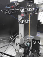
Conventionally used spontaneous Raman scattering is extremely weak, so measurement takes a long time. By using nonlinear Raman scattering, we are developing high-speed imaging methods that distinguish tissues and lesions without staining.
A browser that supports the video tag is required to play the video.
Left: nonlinear Raman imaging of adipocyte-like cells observed at 100 frame/s, faster than video rate. Right: rigid endoscope using nonlinear Raman scattering.
Development of synchronized ultrashort pulse lasers
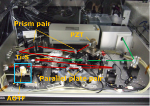 Nonlinear Raman scattering imaging requires special light sources. The sample must be irradiated simultaneously with two laser pulses of different wavelengths, each with a narrow spectral bandwidth and a short (picosecond) duration. In addition, the wavelength of one of the lasers must be tuned at high speed. By developing such laser light sources ourselves, we aim to realize label-free imaging that can be used in clinical settings.
Optical transillumination imaging
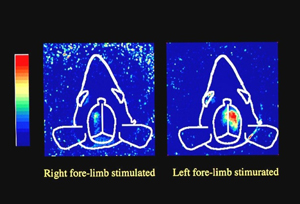
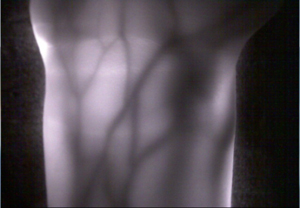
Optical biomedical measurement is expected to become a new diagnostic technology because it enables measurement not only of the morphology of internal organs but also of vital functions. Among these techniques, transillumination imaging can be realized with very simple equipment. We are studying transillumination imaging of functional changes in the brain and organs of laboratory animals, and transillumination imaging of blood vessels to support treatments such as intravenous injection in people whose veins are hard to see, and for vein authentication to strengthen information security in the future.
|
Transillumination imaging of rat brain function |
Transillumination imaging of blood vessels in the adult forearm |
Fluorescence imaging
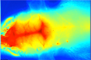
Fluorescent agents that selectively accumulate in cancer and fluorescent agents for angiography have been developed. Using them, we are working on fluorescence imaging of in vivo functional information. We are also developing image-processing methods to restore the blurring of fluorescence from deep inside the body.
Cerebral fluorescence angiography in the rat