To the laboratory website
Outline
The goal of this laboratory is to develop new technologies and approaches in the field of biological science and biomedical applications by applying physics and electronics technologies to biomedical problems. Specifically, we are conducting the research on electron paramagnetic resonance (EPR) imaging to visualize specific molecules in vivo (molecules called free radicals). Through such research, we aim to develop new approaches in the engineering, physics, chemistry and biomedical fields.
Research
We are developing cutting-edge technologies using EPR molecular imaging to visualize functional information in vivo. We also develop measurement technologies to speed up EPR spectrometers and enhance their sensitivity, resolution and stability, establishing advanced applications for ERP imaging.
Development of fast EPR imaging
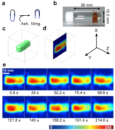
We have developed technology to run EPR imaging (which specifically visualize molecules with an unpaired electron [free radicals]), 10 times faster than before. A uniformly distributed projection method and fast magnetic field sweep were used to speed up EPR imaging. This imaging technology helps to clarify the in vivo behavior of short-lifetime free radicals, thus contributing to clarify disease and life phenomena involving free radicals.
Development of super-resolution EPR imaging
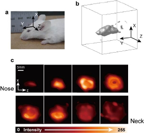
We have developed a resolution-recovery method that significantly enhances the spatial resolution of EPR imaging. This dramatically enhanced resolution allows high-definition in vivo visualization of free radicals. Our aim was to solve the long-standing problem of the limited resolution of EPR image processing.
Development of the simultaneous imaging of isotopically labeled free radicals
Some molecules that serve as medicines have a non-superimposable mirror image in their atomic configuration, although they have the same physicochemical properties. They are called chiral molecules, and this feature is called chirality. We are conducting the development of both a method and the instruments to distinguish chiral molecules that look the same in the body. This research is characterized by labeling of isotopic nitrogen that behaves differently to unpaired electrons in chiral molecules, which are hard to distinguish.

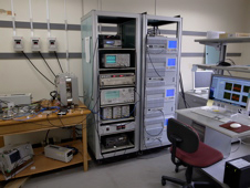
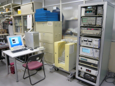
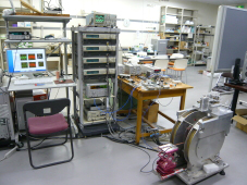
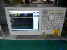
Examples of research equipment
Knowledge and technologies
Basic technologies for research and development related to magnetic resonance spectroscopy
- Microwave circuit technology
- Microwave measurement technology
Technologies and knowledge necessary for the research and development of magnetic resonance molecular imaging
- Basic knowledge of nuclear magnetic resonance and electron spin resonance
- Measurement control technology
- Signal processing technology
Simulation/analysis technologies
- Imaging experiments with a microwave resonator, microwave spectrometer and electron spin are simulated and used in the development of measurement instruments and research on imaging methods. We write most computational codes ourselves.
Keywords
Magnetic resonance molecular imaging, free radical molecules, microwave measurement, image reconstruction, functional imaging
Members
- Professor: Hiroshi Hirata
- Associate Professor: Shingo Matsumoto
- Assistant Professor: Ikuya Nishimura
- Secretary: 1
- Doctoral student: 1; master’s students: 6; undergraduates: 5
Contact
Address: M Bldg, Graduate School of Information Science and Technology, Hokkaido University Kita 14-jo, Nishi 9-chome, Kita-ku, Sapporo, 060-0814
Professor Hiroshi Hirata Tel. 011-706-6762