To the laboratory website
Outline
X-rays are widely used in many areas that are closely related to people’s lives, including radiography/X-ray CTs at hospitals, baggage inspections at airports and non-destructive testing for metal. X-rays, which have high penetrating power, are suitable for imaging deep inside thick samples. If samples have ordered atomic/molecular structures such as crystals and fibers, X-rays can be used to visualize the atomic world. It is well known that the double-helical structure of DNA was determined based on data from X-ray diffraction experiments. Today, research using X-rays is actively being conducted on the atomic structure of proteins that constitute living organisms, significantly contributing to drug discovery.
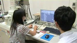
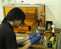
At our laboratory, research using X-rays is conducted on imaging of new worlds that have been inaccessible to date. For our research, we use facilities Japan boasts to the world, such as SPring-8, a large-scale synchrotron radiation facility, and SACLA, an X-ray free-electron laser facility. Such research facilities are found in only a few places worldwide, where X-rays with beautiful waves (coherent X-rays) can be used. At Hokkaido University, we make full use of nanofabrication/measurement systems available at the Open Facility to develop samples and measurement devices. We also use sophisticated computing technology to perform simulations and analyze experimental data.
Resaerch
Nanostructure analysis of biological samples using x-ray diffraction microscopy
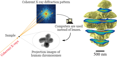
An X-ray diffraction experiment performed with crystallized proteins enables atomic-level ultrahigh-resolution structural analysis. Then, is ultrahigh-resolution structural analysis possible with samples that cannot be crystallized such as cells?
The way to open up this possibility is X-ray diffraction microscopy using X-rays with beautiful waves (coherent X-rays). In X-ray diffraction microscopy, computers are used instead of lenses to obtain images of samples. Since X-rays are highly excellent in penetrating power, they enable the non-destructive observation of cell interior, which is difficult for other microscopes including transmission electron microscopes.
Development of ultrafast coherent X-ray imaging
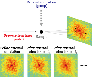
Ultrafast cameras are necessary to observe fast-moving atoms and the nano world. Taking a snap without blurring becomes possible by illuminating an object with a flash that has a shorter light-emitting time than the object’s motion time. X-ray free-electron lasers, which have recently been developed, have a light-emitting time in the femtosecond (10–15 sec.) order, which is shorter than the molecular motion time scale. We illuminate samples with an X-ray free-electron laser, record a time series of snapshots of scattered X-ray patterns, and process them on a computer to take moving pictures of the atomic/molecular world.
Ultrafast cameras are necessary to observe fast-moving atoms and the nano world. Taking a snap without blurring becomes possible by illuminating an object with a flash that has a shorter light-emitting time than the object’s motion time. X-ray free-electron lasers, which have recently been developed, have a light-emitting time in the femtosecond (10–15 sec.) order, which is shorter than the molecular motion time scale. We illuminate samples with an X-ray free-electron laser, record a time series of snapshots of scattered X-ray patterns, and process them on a computer to take moving pictures of the atomic/molecular world.
Examples of X-ray facilities used for research
Examples of instruments at the Open Facility of Hokkaido University used for our research
- Laser direct lithography system
- Plasma CVD system
- Reactive ion etching system
- Field-emission-type scanning electron microscope
- Ramam microscope system
- Color 3D laser microscope
- Scanning probe microscope
- X-ray diffraction system
Examples of research equipment in the laboratory
- Rotating cathode X-ray generation system
- Nanocrystal fabrication system
- He-Ne laser coherent imaging system
- Linux server
Knowledge and technologies
Technologies and knowledge necessary for X-ray-related research
Advanced X-rays such as synchrotron radiation and X-ray free-electron lasers are used for measurement. X-ray generators in the laboratory and the Open Facility are also used. Students can learn X-ray science and optics through research. For measurement, a broad range of samples are used, which allows students to acquire extensive knowledge in biology, medicine and material science. Students can also learn a wide range of technologies concerning measurement and instrument control through experiments.
Technologies and knowledge necessary for laser-related research
Femtosecond-pulsed lasers are used for time-resolved measurement. For the development of coherent X-ray imaging methods, a visible-light laser in the laboratory is used for experiments to verify principles.
Technologies and knowledge necessary for nanotechnology-related research
Photolithography, CVD and other technologies are used to fabricate micro-devices. Various types of microscopes are also used to observe these devices. Through experiments, students can acquire a wide range of technological skills concerning a large variety of nanotechnology-related advanced instruments at the Open Facility of Hokkaido University.
Technologies and knowledge necessary for data analysis and instrument control
Software necessary for data analysis and instrument control is developed by ourselves by programming using C, Python, Matlab, LabVIEW, etc. A Linux server and GPU are also used for the development. Students can acquire technologies concerning scientific computation and instrument-control programming.
Technologies and knowledge necessary for instrument design
Creo Elements/Pro, Vectorworks, and other CAD software are used for instrument design.
People who are interested in these key words, please visit this laboratory.
Bioimaging, ultrafast imaging, coherent X-rays, synchrotron radiation, X-ray free-electron lasers, femtosecond pulsed lasers, computer programming, nanofabrication, nanoimaging, photolithography, CVD, instrument control, instrument design
Members
- Professor: Yoshinori Nishino
- Assistant Professors: Marcus Newton, Takashi Kimura
- Undergraduates: 3
Contact
Address: 04-301, Creative Research Building
Kita 21-jo, Nishi 10-chome, Kita-ku, Sapporo, Hokkaido, 001-0021
Tel.: 011-706-9354
Fax: 011-706-9355